Total Visioncare Examination - The best way to look after your eyes
Your sight is important, so you should look after it with the best eye test possible. A First Tier Total Visioncare Examination is the most thorough eye examination possible and the best way to care for your eyes. There are very few Optometry practices in the UK that have the expertise and equipment to be able to carry out such an examination for you. This ophthalmic examination provides early detection of many eye conditions, including glaucoma, macular degeneration, cataracts and visual field problems. If we detect any of these conditions, we can initiate treatment early. If your eyes are healthy, then these tests provide a perfect baseline for the health of your eyes which means if you have any minor problems or changes in the future, we can find these immediately.
In addition to your routine eye examination, we use the latest technology to carry out extra tests. You will have your eyes 4D scanned inside and out. We will measure your corneal thicknesses, hospital standard visual field testing, and pressure measurements. We assess for binocular vision and muscle problems, dry eye and lid problems. We recommend the Total Visioncare Examination to all our adult patients. The examinations last for approximately one and a half hours. Following the tests, we will discuss the results with you so that you can leave knowing your eyes have had the very best examination possible, allowing detection and prevention of sight-threatening problems. When you book your next eye exam, ask for the Total Visioncare Examination. Call to book today on 01634 757227.
Do I need an hour and a half long eye examination which scans the nerves and tiny cells at the back of my eyes?
Yes, of course, because it is your eyesight! It’s also the eye examination we carry out for all our friends and family.
Why? It can prevent you from losing your eyesight. During the Total Visioncare Examination, we assess your risk for several conditions, including glaucoma, diabetic eye disease, macular degeneration, cataracts, and much more. Finding eye conditions early can slow any deterioration and sometimes prevent them from occurring. We service and MOT our cars yearly. You only have one set of eyes; having a very thorough check-up every year or two is the only sensible way to ensure they keep working efficiently. Many eye conditions do not have symptoms in the earliest stages, but if we find them early enough, most can be treated successfully.
Why is the Total Visioncare Examination better than a standard eye test which seems pretty thorough to me?
Although our standard eye examination is longer and more thorough than the majority of high street eye tests, without using advanced OCT 4D eye scans, we just cannot detect eye diseases as early. By using the non-invasive light to examine your eyes, we can see the individual layers at the back of your eyes (some are only one cell thick). This means we can spot the tiniest sign of damage long before the conventional methods of looking in your eyes. Using the scans and techniques in the Total Visioncare Examination, we can detect some eye conditions up to 8 years earlier than usual.
Why doesn’t every optician offer this?
The OCT eye scanner and other equipment used during the examination cost hundreds of thousands of pounds, which means that only a handful of opticians in the UK have the kit to offer this level of eye screening.
Do the scans use lasers? Is this safe?
An OCT (optical coherence tomography) scan is entirely different from the routine eye photos taken by many opticians or your diabetic clinics. Using safe, low-powered lasers, the OCT instrument scans below the eye’s surface. Unlike most types of lasers, this machine uses an entirely safe wavelength of light to take the images. You can have thousands of scans with no risk to your eye health. There are no x-rays involved. It is completely painless, non-invasive and takes only a matter of seconds. Your results are available instantaneously, and it is an excellent way for you and your optometrist to understand better the health of your eyes or any eye conditions. You can find out more about how the scans work here.
If I haven’t got an eye problem, are the additional scans and tests necessary?
Hopefully, we won’t find any problems with your eyes, but the scans still act as a perfect baseline measurement for future reference. Many eye conditions are diagnosed by ‘changes’ to your eyes. If we can measure every tiny part of your eye today down to 1 micrometre, we can spot the smallest changes so quickly in the future. Everyone’s eyes are different (a bit like a fingerprint), and until we know what is normal for you, it can make change tough to detect.
I can see well, and my eyes don’t hurt, so surely there’s no problem.
Wrong. The vast majority of eye conditions we diagnose have no symptoms in the early stages, including glaucoma, cataracts and macular degeneration.
I don’t have a family history of glaucoma, so do I need to worry?
Yes. Although the condition is more common if you have a family history, anyone can suffer from this sight-threatening condition. Age is one of the most significant risk factors for glaucoma. Approximately 2% of the population over 40 years old has glaucoma. It is estimated that by 80 years old, as many as one in ten people may have the condition.
Glaucoma is often called the thief of sight as it has no symptoms until it is well established, often creeping up and stealing your sight before you are even aware there is a problem. When symptoms become noticeable, it's usually only when significant loss of vision (up to 40%) has occurred. Therefore, everyone must understand the need for regular advanced glaucoma eye examinations, which can detect the condition long before you can and stop you needlessly losing your sight.
Over 90% of individuals diagnosed early will retain useful sight for life. Despite such promising results, it is estimated that over 50% of cases of Glaucoma remain undetected; that’s 300,000 people who are needlessly losing their sight just because they have neglected to have their eyes tested properly. If we detect glaucoma during your Total Visioncare Examination, then treatment can be started to stop you from losing any sight.
Why is smoking relevant to my eyes?
If you have smoked at any point in the past, you are 42 % more likely to get cataracts and are also at increased risk of glaucoma. Smokers are up to four times more likely than non-smokers to develop age-related macular degeneration (AMD), and smokers with a genetic predisposition to AMD are up to 20 times more likely to get the condition. Macular degeneration is the leading cause of sight loss in the UK. Smoking may as much as double the risk of developing diabetes which can lead to diabetic retinopathy and sight loss. If we find any of these conditions early enough, the prognosis can be significantly improved.
What examinations can I expect?
Please read on for further information about some of the additional tests which we may include in your Total Visioncare Examination.
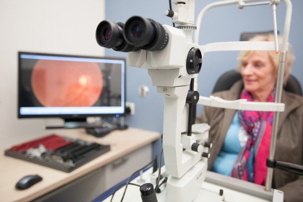
Heidelberg multicolour Optical Coherence Tomographer Spectralis 4D eye scan
We are proud to be one of the first practices in the UK to have the new Spectralis 4D OCT (Optical Coherence Tomographer). Using this phenomenal machine and incredible computer technology, we can now diagnose many more eye problems at the earliest possible stage. We can treat eye conditions sooner and prevent many from even developing at all, which is the main aim of our job.
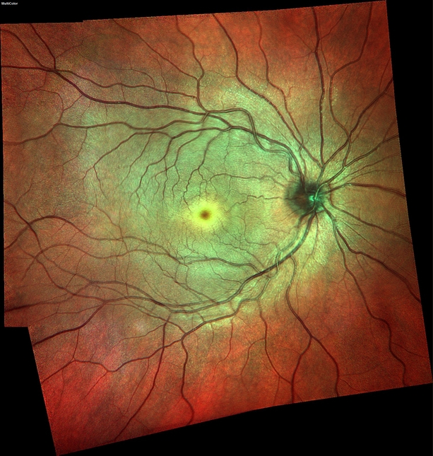 The Retina is the inner lining at the back of your eye. It acts similarly to the film in a camera, capturing the image of what you see and then transferring this image to the brain for interpretation. When an Optometrist looks into your eye or takes a photograph, it is the retina they are looking at.
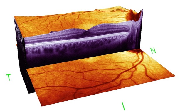
Conventional digital cameras, used by many opticians, just look at the top surface of the retina, but many eye problems start at a deeper level. The Spectralis 4D OCT Eye Scanner is equivalent to a CAT scan of your eye. Unlike retinal photographs, this new system not only allows us to take a very high-resolution detailed image of your retina but also helps us look below the surface at each layer of the retina to create a High-Definition map that provides us with information never before available. We can even see details in the walls of your blood vessels and tiny individual nerve fibres. This helps us to find the slightest sign of problems such as diabetic eye disease, vascular problems, neavi (moles), glaucoma, macular degeneration, oedema and retinal holes long before they may be visible on the retinal surface. Also, long before they can be seen by standard retinal photography or by looking into your eyes (ophthalmoscopy). The new Spectralis 4D OCT can help us diagnose and monitor all these conditions with the accuracy of one-thousandth of a millimetre. Earlier detection leads to earlier treatment, a better prognosis and, in some cases, prevention of conditions developing at all.
 The Spectralis 4D OCT scan not only helps us detect eye conditions but can sometimes also show signs associated with brain tumours, high cholesterol or uncontrolled blood pressure, thus saving further health problems in the future and may even help prevent a stroke or heart attack.
We recommend Spectralis 4D OCT scanning for all patients as it acts as not only a very thorough assessment of your eye health but also an excellent baseline for future comparison to help diagnose any changes at the earliest signs. Scanning is particularly advisable for anyone diagnosed with cataracts, macular degeneration, glaucoma, diabetes or with a family history of any of these conditions. These scans go far beyond the measurements normally carried out in the hospital and, therefore, can allow more accurate monitoring of your condition.
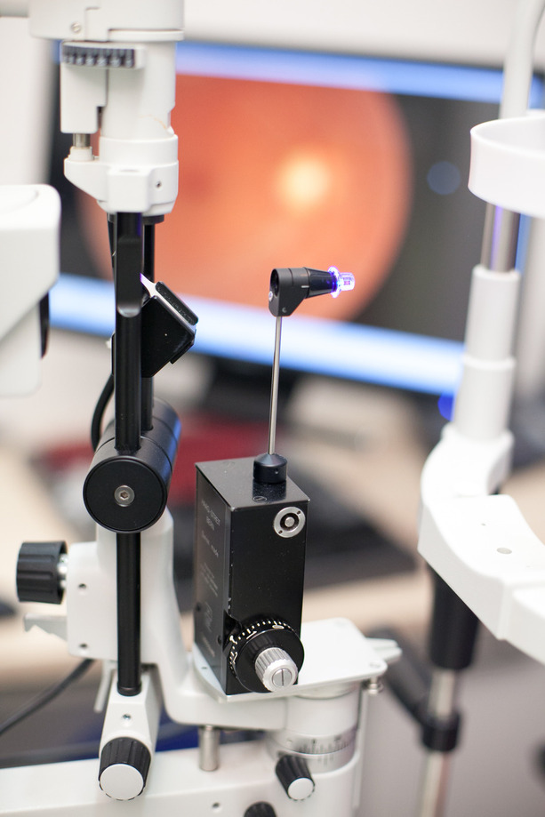
Heidelberg Spectralis 4D Optical Coherence Tomographer eye scanner uses a very safe low-powered imaging laser to accurately measure the thickness of the nerve fibre layer. It is a non-invasive technique and doesn’t use any harmful rays like x-rays.
The process requires you to place your chin on a rest, much like many of our other instruments and look at a fixation light. Unlike the typical cameras most opticians use, we get a very sharp, clear image without dilating your pupils and even through cataracts. Each scan takes just a few seconds on each eye. It does not touch your eye and requires no drops so that you can drive straight after your appointment.
After the images have been viewed and assessed, they are stored electronically for your next visit, where referring to the previous images, we can spot even tiny changes (1 micrometre!). Allowing for an easier and more accurate method of monitoring the health of the eyes year after year. If necessary, the retinal images can be passed on to GPs or Hospitals for their reference too. Spectralis 4D OCT eye scanning is recommended for all adult patients and can be used at any age if advised by your Optometrist.
The 4D Spectralis OCT also has a unique role in macular imaging and glaucoma detection.
We recommend advanced glaucoma screening for everyone over 35. Book your appointment today and prevent sight loss. 01634 757227.
Humphrey Threshold Visual Field Analysis
Your field of vision is every area you can see with your eyes pointing straight ahead. It is essential to have a wide field of vision to help us navigate around, especially when walking or driving. You can lose part or all of your visual field in many different ways, including cataracts, glaucoma, strokes, circulatory disorders, neurological conditions, certain brain tumours and even smoking. It is essential to have your visual field regularly assessed as many conditions, if caught early, can be treated.
Our Humphrey Visual Field Analyser is considered the gold standard in advanced visual field testing (and is the 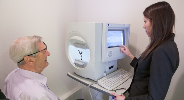 Instrument recommended by Ophthalmologists). It accurately assesses how wide and sensitive your visual field is. Unlike basic visual field screening, which concentrates on evaluating large chunks of your vision at a time, the Humphrey Visual Field Analyser can pick up even very subtle defects in your visual field by analysing each nerve pathway in detail. This allows for the detection of problems. However, even a perfect visual field should regularly be measured as this provides an accurate individual baseline to monitor future changes again, allowing earlier diagnosis of problems.
 Advanced pressure testing
A Goldmann pressure test can be conducted to accurately test and monitor the pressure inside your eye; this is considered the “gold standard” test. This is the best and most comparable way of monitoring your eye pressures. This test does not require the puff of air used by many pressure machines.
Pachymetry
Pachymetry uses a small instrument to accurately measure the thickness of your cornea- the clear window at the front of your eye. The eye pressure measured with the puff of air or Goldmann tonometer is influenced by the thickness of your corneas. Thick corneas can mean your exact eye pressure is less than we have measured, and thin corneas lead to an under-reading. Using the Pachymeter, we apply an adjustment to your intraocular pressure measurement to more accurately assess and monitor the pressure in your eye. This test often prevents unnecessary hospital referrals as well as identifying patients with apparently low pressures who may be at risk of glaucoma.
The test takes just a few seconds and is painless.
 Slit Lamp Biomicroscopy
To accurately view the interior and exterior of the eye in 3D, we use slit lamp indirect biomicroscopy - a technique which involves specialist skills and an advanced piece of equipment to view all the hard-to-reach corners of the eye. This method can help us identify retinal bleeds, holes, tears and detachments far more accurately than traditional methods of looking in your eyes. Biomicroscopy also aids detection of diabetic eye disease, glaucoma, Papilloedema (pressure behind the eye), Truemacular degeneration, hypertension and many other conditions.
Colour Vision
We check colour vision firstly in childhood to establish if there is an inherited problem and gain a baseline for future reference. Some colour deficiencies that occur later in life can be caused by illness, accident or poisoning. Colour vision is easy to check. The test is performed under daylight lighting conditions in the consulting room. There are several variations of colour vision testing, some requested for particular occupations.
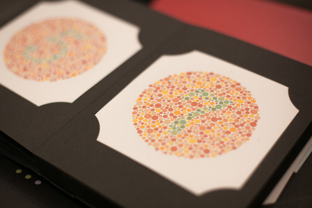
Colour vision deficiency is commonly known as colour blindness. Real colour blindness when people see things only in greyscale is very rare. Most people with colour vision problems have an inherited red-green colour deficiency which leads to confusion of colours in that spectrum. Due to its inheritance pattern, it is most common in men. Approximately 8% of men and 1% of women are colour deficient. There are other types of colour deficiency that can affect other colour groups as well. Red, green and blue cone cells at the back of our eyes detect colour variations. In colour deficient eyes, one or more types of the cones may be absent or deficient, affecting the ability to perceive certain colours.
Binocular vision
It is the ability of the two eyes to work as a pair. Binocular vision allows us to perceive depth and maintain a single image, i.e. avoiding double vision. Binocular vision problems can often be subtle and lead to signs of visual stress such as:
- Print jumping as you read
- Difficulty focusing on individual words or letters.
- Diplopia (double vision)
- Tired eyes when performing visual tasks
- Strabismus (squint)
Your binocular visual balance will be assessed at every eye examination to ensure you get the most comfortable vision possible, eliminating signs of visual stress. If a binocular vision problem is identified, there are many methods we can use to help alleviate symptoms, including using prismatic correction in spectacle lenses and eye exercises.
Binocular vision problems may be present from childhood or develop later in life. Certain conditions may lead to sudden binocular vision problems; these include diabetes, hypertension and nerve palsies.
What does it cost?
At Buchanan Optometrists, we are transparent in our pricing. We use the very latest equipment that is not even available within private or NHS hospitals locally. Your comprehensive ophthalmic examination takes much longer than a routine sight test carried out by most opticians. Our consultant optometrists are exceptionally trained to interpret and diagnose any conditions that may be apparent from your results. This is the best way to look after your eyes to ensure your continued health and optical comfort. The cost is only £390
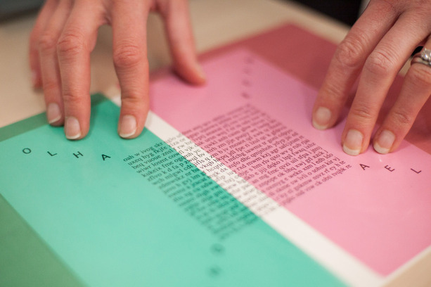
Eye Tracking and Coloured overlay assessment
This assessment is not included in the Total Visioncare Examination but can be carried out for adults and children following their eye examination.
Coloured overlays and spectacles have been found to have beneficial effects for sufferers of both visual stress and reading difficulties, including many forms of dyslexia. Following a comprehensive eye test to rule out any prescriptive or ocular motor problems, an eye-tracking assessment with one of our specialist optometrists will conclude if a coloured overlay or spectacles can help you or your child reduce visual stress, increase reading speed, and improve concentration.
Symptoms and signs of visual stress may include:
- Discomfort when reading
- Glare from a white page
- Seeing Patterns in the print
- Migraines
- Blurring of print
- Letters and words appear to be moving
- Re-reading the same line frequently
- Moving the book a lot to focus
- Skipping or missing words
- Poor concentration when reading
Coloured Overlay Assessment and Eye Tracking are available for six years old upwards.
Read more about eye tracking and coloured overlays here.
Book your appointment today on 01634 757227 or click HERE and we will call you straight back!